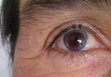
Cell implants, genetic therapy, even opto-genetics are making headway in clinical trials and eveb treatment various forms of blindness.

Cell implants, genetic therapy, even opto-genetics are making headway in clinical trials and eveb treatment various forms of blindness

Recently, Scientists have accomplished what formerly was conserved for miracle workers: they have provided blind people the ability to see much better. In 2017, the vision field saw a massive advancement with the approval Luxturna, the initial genetics therapy to fix vision loss in specific patients with childhood years onset blindness.

And simply recently, researchers reported that a retinal implant enabled a 69-year-old woman with macular degeneration to more than double the number of letters she might determine on a vision chart.

Macular Degeneration of the Eye- Lasik and Cataract Surgery Los Angeles

” It’s early information however extremely promising, consisting of one patient with excellent vision gains, for an illness where we don’t have any type of treatment alternatives,” claims Thomas Albini of the University of Miami’s Bascom Palmer Eye Institute who was not involved in the research study.

The implant, offered to five patients with dry age-related macular degeneration (AMD), is a single sheet of retinal pigment epithelial (RPE) cells originated from human embryonic stem cells. Various other groups across the globe are developing their very own type of RPE implants, and this type of approach is simply among a plethora of modalities being evaluated to either decrease or reverse various forms of blindness.

Up until now, the regenerative medicine field has actually identified exactly how to generate RPE cells, but various other cell types have proved more difficult “Getting photoreceptors or various other neurons inside the retina achieving which has been a golden target. No one has been successful in generating these cells,” says Jean Bennett, a professor of ophthalmology at the University of Pennsylvania.

Scientists have nonetheless developed creative technologies for sight restoration to deal with a variety of blindness conditions, some of which are enumerated below:

Cell implants

In the most recent advancement on dry AMD, published in Science Translational Medicine, a team of scientists and also engineers at the University of Southern California (USC) in Los Angeles initially evolved human embryonic stem cells (ESCs) into RPE cells – those that develop a monolayer that supports the health and wellness of the overlapping photoreceptor-harbouring retina. When spread out on an itsy-bitsy polymer platform (simply 6 mm by 3 mm), the cells assembled into a polarized mono-layer, just like that found in the human eye.

“What we have is a replica of the cells that are missing, because of degeneration, in patients with advanced dry AMD,” says Amir Kashani, a clinical scientist who performed the surgeries.

To insert the cells, Kashani made a one-millimetre incision in the retina, and with the implant folded in fifty percent “like a taco,” he moved it into the eye and unfolded it underneath the retina– which holds the patch in place – on top of the damaged area.

Three patients saw enhancements within three months of receiving the implant, including one patient who could check out additional letters on a vision chart. The other two patients had improvement in fixation, the capability to concentrate one’s gaze on an object.

Post-operative photographs of one subject 180 days (E) and another subject 120 days (F) after implantation. The bio engineered implant (black dotted line) is located near the area where vision cells have actually died (white dotted line).

The team is currently registering an additional 15 patients onto this trial and is planning a larger, phase 3 trial, additionally in dry AMD patients. “The objective is to try to fix much less severe disease so there is even more of a possibility for visual improvement,” says Kashani.

One more recent stem cell-based tissue approach from scientists in the United Kingdom resulted in vision improvement in two older patients with the wet form of AMD. In these cases, blood vessels in the eye grow abnormally and can lead to bleeding, the destruction of RPE cells, and eventual blindness. The developers also used a coated, synthetic membrane on which they assembled RPE cells differentiated from human ESCs and surgically delivered the eye spot. Two patients, a woman in her 60s and a man in his 80s, obtained the ability to read an additional 29 and 21 letters, respectively, on an analysis chart through a year. Each patient additionally had astonishing improvements in reading speed, from 1.7 to 82.8 and from 0 to 47.8 words per minute, respectively.

The initial presentation that human ESCs differentiated into RPE cells are safe was in 2012, when Steven Schwartz of the University of California, Los Angeles, and his colleagues effectively infused ESC -derived RPE cells into two legally blind patients, one with dry AMD and another with Stargardt’s macular dystrophy, one more degenerative eye disorder. In a 2014 follow up report of 18 patients, the team along with the manufacturer of the cells, Astellas Pharma, showed that 10 of the patients had some vision improvement. Schwartz and his colleagues are currently planning to register a bigger trial to evaluate if injecting a higher number of human ESC – derived RPE cells than used in the initial trial will certainly be efficacious.

One concern with using human ESCs is a prospective immune reaction, and subsequent rejection of the cells as foreign. To establish customized cell therapies originated from patients’ own cells, a group in Japan led by Masayo Takahashi at the RIKEN Centre for Developmental Biology have relied on reprogramming cells from skin fibroblasts into induced pluripotent stem (IPS) cells and then right into RPE cells. Her team recently revealed that a wet AMD patient who had a sheet of such RPE cells implanted into the eye did not have additional vision atrophy throughout a year.

There are likewise researchers working on a much more scalable approach, using donated, immune-matched IPS cells to treat several patients. A bank of IPS cells from a variety of donors would certainly be much more feasible, affordable + friendly, and also hassle-free than making IPS cells from each patient’s own cells.

Gene therapy and devices

To fix specific genetic defects associated with blindness disorders researchers have relied on gene therapy—and have achieved striking results. Luxturna, the gene therapy that FDA approved in 2015, fixes a mutation discovered in Leber congenital amaurosis (LCA) – a rare acquired form of childhood – and various other types of genetic retinal dystrophies harbouring a mutation in the RPE65 gene. The protein inscribed by the gene is an enzyme required for retinal cells to produce visual pigments. These inherited disorders generally start with night blindness and can proceed to a degeneration of peripheral vision and then total vision loss in childhood years.

One goal is to confer light level of sensitivity to retinal cells that usually do not have the capability to sense light.

An additional genetics therapy, still in the works, produces a healthy protein that obstructs the activity of vascular endothelial growth factor (VEGF) and has currently been shown to be safe and effective in an early-stage wet AMD clinical test.

A twist on gene therapy is an optogenetic approach in which a vector encoding a channel rhodopsin from environment-friendly algae is provided to retinal ganglion cells. The company Allergan is currently evaluating the strategy, developed by Zhuo-Hua Pan, a vision researcher at Wayne State University, in a beginning trial for patients with retinitis pigmentosa, in which patients lose light-sensing retinal cells. The objective, according to Pan, is to confer light sensitivity to retinal cells that usually don’t have the capability to sense light. While some genetics treatments, such as the one approved for LCA, target specific mutated genetics, optogenetic therapy might be utilized to deal with any type of blindness due to a loss of photoreceptor cells, says Pan.

Unproven stem cell techniques

The genetics and cell therapies in development are still in the early stages, and it’s not yet clear from the small numbers of patients dealt with up until now exactly how extensively appropriate they might be to people with vision loss. As cautious scientific research on these technologies inch their method towards ever-larger trials, there are centres offering unapproved treatments to customers with blindness – in spite of there being no stem cell – based therapies authorized for severe eye diseases.

Just recently, three women, each with macular degeneration, received an unverified stem cell treatment in both eyes in a clinic in Florida. All three were permanently blinded. Albini says the clinic’s protocol was a warning they weren’t operating very carefully. “The treatment ought to be performed in just one eye,” he says, whereas the stem cell clinic in Florida was providing its therapy to both eyes, blinding the females bilaterally.

An additional warning: the business model. “It’s important for patients to comprehend that legitimate clinical tests do not charge patients for the therapy,” adds Schwartz.

Regardless of the negative Public Relations from rogue players, ophthalmology researchers are bullish on what new treatments and advancements the following decades will certainly bring. Says Bennett, “It’s an exciting time. The vision is actually flourishing”.
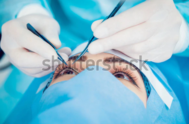
CATRACT SURGERY

Los Angeles Beverly Hills Painfree Lasik Cataract Surgery – Khannainstitute.com

At some point after age 50, the majority of us are likely to hear our ophthalmologist say, “You have cataracts.”

A cataract is a clouding of the lens inside the eye, causing vision loss that cannot be corrected with glasses, contact lenses or corneal refractive surgical procedure like LASIK.

As frightening as cataracts might sound, contemporary cataract surgical treatment usually can recover vision lost to cataracts– and an minimize your dependency on eyeglasses too.

A lot of cataracts are related to the aging process and are common amongst older Americans.

As well as the ocurrence of cataracts in the United States is expected to grow substantially in the years ahead, due partially to the aging of the population. It is expected that 50.2 million Americans will have cataracts by the year 2050, according to NEI.

Fortunately, modern cataract surgical treatment is one of the safest and most effective surgical procedures executed today.

Greater than 3 million cataract surgeries are performed in the USA every year, with the vast majority of these treatments generate exceptional visual results.

Cataract Surgical Treatment Fundamentals

In cataract surgical treatment, the lens inside your eye that has actually become cloudy is eliminated and replaced with an artificial lens (called an intraocular lens, or IOL) to restore clear vision.

The treatment typically is carried out on an outpatient basis and also does not require an overnight stay in a hospital or various other treatment centre.

The majority of modern cataract procedures include the use of a high-frequency ultrasound device that separates the cloudy lens into small pieces, which are then carefully removed from the eye with suction.

This procedure, called phacoemulsification or “phaco,” can be performed with smaller incisions than previous surgical methods for cataract removal, promoting faster healing and minimizing the risk of cataract surgical treatment difficulties, such as a retinal detachment.

Nevertheless, remnants of the cloudy lens have been removed from your eye, the cataract surgeon inserts a clear intraocular lens, positioning it securely behind the iris and pupil, in the same location your natural lens inhabited. (In special cases, an IOL may be placed before the iris and pupil, however this is much less common).

The surgeon then completes the cataract removal and IOL implantation procedure by closing the incision in your eye (a stitch may or may not be required), and also a safety shield is placed over the eye to keep it risk-free in the early stages of your cataract surgery recovery.

Laser Cataract Surgery.

Recently, a number of femtosecond lasers – similar to the lasers used to create the corneal flap in all-laser LASIK– have been approved by the FDA for usage in cataract surgical treatment performed in the United States.

These lasers have acquired approval for complying with steps in cataract surgical treatment, minimizing the need for surgical blades and various other hand-held tools. They are used for:

1. Creating corneal incisions to permit the surgeon accessibility to the lens.

2. Removing the anterior capsule of the lens.

3. Fragmenting the cataract (so much less phaco energy is required to break it up and remove it).

4. Creating peripheral corneal incisions to reduce the astigmatism (when needed).

Laser cataract surgical treatment (or, much more accurately, laser-assisted cataract surgery) is relatively brand-new and substantially enhances cataract surgical treatment cost, mainly because the laser can cost from $300,000 to $500,000 for a surgeon to purchase and also there are various other substantial costs associated with the usage and maintenance of this technology.

While researches have revealed that lasers can improve accuracy throughout specific actions of cataract surgery, they may not necessarily improve cataract surgical treatment safety and security, recovery time and also visual results in every situation.

For the most recent information about laser cataract surgery, ask your ophthalmologist throughout your preoperative eye examination and cataract surgical treatment assessment.

Planning for Cataract Surgery and Choosing An IOL.

Before cataract surgery, your optometrist and/or ophthalmologist will perform a comprehensive eye examination to examine the overall health of your eyes, assess whether there are reasons why you ought to not have surgical treatment and identify any type of risk factors you might have.

A refraction additionally will be performed and carried out precisely identify the amount of near-sightedness, farsightedness and/or astigmatism you have prior to surgery. Additional measurements of your eyes will certainly be taken to determine the curvature of your cornea and also the length of your eye.

These dimensions are necessary to help your cataract surgeon choose and pick the appropriate power of the intraocular lens and give you the best vision possible after surgery.

Today you have many types of IOLs to choose from for your cataract surgery, depending on your particular needs. In addition to IOLs that correct near-sightedness as well as farsightedness, there are now toric IOLs that correct astigmatism as well.

If you do not mind putting on glasses after cataract surgical treatment, a mono-focal lens implant usually is used. Frequently, only part-time use of reading glasses is needed after cataract surgery with mono-focal IOLs. But if prescription eyeglasses are needed which often is the case if you only need cataract surgical treatment in one eye, your ophthalmologist typically will recommend brand-new glasses for you around one month after surgery.

If you like the concept of being much less dependent on glasses after cataract surgery, one way to correct presbyopia and minimize your need for reading glasses is to have your cataract surgeon  adjust the power of your mono-focal IOLs (assuming you have cataract surgery performed executed in both eyes) to provide you a monovision correction, similar to monovision with contact lenses.

Another option is to choose among a variety of advanced presbyopia-correcting IOLs to improve your reading vision without compromising your distance vision. Presbyopia-correcting IOLs include accommodating IOLs and multifocal IOLs; both types are developed to provide a better array of vision after cataract surgical procedure than traditional mono-focal IOLs.

Realize that not everyone is an excellent prospect for these premium IOLs, and choosing a presbyopia-correcting IOL will enhance the out-of-pocket cost of your cataract surgery, since the included cost of these advanced lens implants is not covered by Medicare or various other insurance policy strategies.

Prior to cataract surgical procedure, in addition to discussing the various kinds of IOLs, you will be advised about what to expect before, throughout and after your procedure. This information which may be presented orally, in composing, using video presentation or a combination of all three – is meant to help you make an enlightened decision regarding whether to proceed with the surgical procedure.

If you have any type of inquiries or concerns regarding cataract surgery, be sure to review them with your ophthalmologist and cataract surgeon before signing “notified consent” papers authorizing surgical treatment.

Additionally, talk with your ophthalmologist about all medicines you are taking, consisting of non-prescription (” over the counter”) solutions and nutritional supplements. Some medications and supplements can increase your risk of cataract surgery complications and might need to be terminated before surgical treatment. Ask your medical professional for details.

Cataract Surgical Treatment Recovery

An uncomplicated Cataract surgery typically lasts only around 15 minutes. However, expect to be at the surgical centre for 90 minutes or longer, due to the fact that extra time is needed to prepare you for surgery (dilating your pupil; administering preoperative medication) and for a quick post-operative evaluation as well as instructions regarding your cataract surgical treatment recovery prior to you leaving.

You should have somebody drive you home after cataract surgery; do not attempt to drive until you have actually seen your ophthalmologist the day after surgery and he or she evaluates your vision as well as confirms that you are safe to drive.

You will certainly be prescribed medicated eye drops to utilize a number of times each day for a few weeks after cataract surgery. You additionally need to wear your protective eye shield while sleeping or napping for about a week after the surgical procedure. To safeguard your eyes from sunlight and other bright light as your eye recovers, you will certainly be given a special set of post-operative sunglasses.

Also, many centres require someone to be with you after cataract surgery if you received anaesthesia. As one has to be sure to ask about this requirement prior to your cataract procedure so you are prepared for surgery day.

While your eye heals, you might experience some eye inflammation as well as blurred vision during the very first few days and even weeks complying with the procedure.

During at least the very first week of your recovery, it is necessary that you avoid:

• Strenuous activity and heavy lifting (absolutely nothing over 25 pounds).

• Bending, exercising and similar tasks that might emphasize your eye while it is healing.

•   Protection from water splashing which could be the cause of eye infection. Maintain your eye closed while showering or bathing. Additionally, avoid swimming or Jacuzzis for a minimum of two weeks.

• Any type of activity that would certainly reveal your recovery eye to dust, grime or various other infection-causing pollutants.

Your cataract surgeon may provide you various other guidelines and recommendations for your cataract surgery healing, depending upon your particular requirements and the result of your treatment. If you have any kind of inquiries at any time after cataract surgery, call your ophthalmologist for recommendations.

If you require cataract surgical treatment in both eyes, your surgeon usually will prefer that you wait one to three weeks in between treatments, so your initial eye has actually recovered completely and also you have excellent vision in that eye prior to when the second surgery is performed.

Eyeglasses After Cataract Surgical Procedure

Unless you pick presbyopia-correcting IOLs, it’s most likely you will certainly require reading glasses after cataract surgery to see near objects clearly. Also, people who choose these premium IOLs often find reading glasses are useful for near vision tasks and seeing extremely fine print.

In case you have some moderate refractive errors present after surgery, you might intend to use spectacles with progressive lenses full time after your surgical treatment to achieve the very best possible vision at whatsoever distances.

Also individuals who have an exceptional visual end result and can see well without glasses after cataract surgical procedure frequently choose to wear glasses permanently after their treatment to safeguard their eyes and due to the fact that they really feel much more like themselves wearing eyeglasses after surgery, if they have used glasses the majority of their life.

If you select to wear glasses after cataract surgery, lenses with anti-reflective covering and photochromic lenses are extremely recommended for the very best vision, convenience and appearance. Ask your eye treatment professional for details and to show these lenses.

Researchers Working with Eye Drops to Dissolve Cataracts

Cataract surgical treatment is confirmed to be risk-free and effective, with a reduced risk of severe difficulties. However, wouldn’t it be excellent if eye drops could dissolve cataracts without the need for expensive surgical treatment? For someone, they might help people who reside in locations without surgeons  or economical healthcare.

In a recent study of congenital cataracts (significance cataracts that exist from birth), University of California San Diego scientists discovered that a particle called lanosterol protects against cataract-causing proteins from sticking to each other, maintaining the human lens clear. However, if the molecule is absent because of a gene mutation, the proteins will adhere and make the lens cloudy.

The researchers developed eye drops containing lanosterol, evaluated them in rabbit eye lenses and discovered the lenses were clearer after a couple of days. In live dogs’ eyes the drops likewise lowered the effect of their cataracts.

The drops do not function as well as surgical treatment, due to the fact that they do not remove the cloudiness in the lens– they just decrease it. Additional screening might generate a much more efficient formula, yet that will certainly take some time.

Still, the prospect exists for cataract-dissolving drops to be made use of regularly one day not simply in pets, however likewise in human beings.

Visual Impairment for Teens

Safety with Khanna Vision Institute

The human eye is like a video camera that accumulates, concentrates, and transmits light through a lens to produce an image of its environments. In a camera, the image is developed on film or an image sensor.  In the eye, the photo is developed on the retina, a slim layer of light-sensitive cells at the rear of the eye.

Like an electronic camera, the human eye regulates the quantity of light that gets in the eye. The iris (the tinted round component of the eye) regulates the quantity of light travelling through the pupil. It closes the pupil in brilliant light and also opens it broader in low light. The cornea is the clear, protective surface area of the eye. It aids concentrate light, as does the lens, which rests simply behind the iris.

When light gets in the eye, the retina alters the light right into nerve signals. The retina after that sends out these signals along the optic nerve (a cord made up of greater than 1,000,000 nerve fibres) to the mind or brain where the signals are read. Without a retina or optic nerve, the eye cannot interact with the mind, making vision difficult.

What Is Visual Disability?

Lots of people have some sort of visual trouble at some time or the other in their lives. Some can no more see things that are far, others that are near, for example they have issues reviewing fine print. These sorts of problems are commonly conveniently treated with spectacles or contact lenses.

Yet when several components of the eye or mind that are required to refine pictures come to be infected or harmed, serious or total failure of vision can take place. In these situations, vision cannot be totally brought back with clinical treatment, surgical treatment, or restorative lenses like eye glasses or contacts.

The American Foundation for the Blind, a non-profit body, estimates that ten million individuals in the US of A are visually impaired. Visual impairment is a term specialists make use of to define any kind of vision loss, whether it’s a person that cannot see in any way at all or a person that has partial vision loss.

Some individuals are completely blind; however, several others have what’s called lawful blindness. They haven’t lost their sight entirely however have actually lost sufficient vision that they would certainly need to stand 20 feet from an object to see it along with someone with twenty-twenty might from 200 feet away.

What Triggers Visual Impairment Problems?

People rarely lose their vision during their teen years. When they do, it’s normally triggered by an injury like getting struck in the eye or head with a baseball or having a vehicle or motorbike mishap.

Some children have hereditary blindness, which implies they are visually impaired at birth, carried in their genes from parents or grandparents. Congenital blindness can be caused due to a number of reasons – it can be inherited as enumerated earlier, for example, or triggered by an infection (like German measles) that’s transmitted from the mother to the developing foetus during pregnancy.

Problems that might create vision loss after birth consist of:

Amblyopia (articulated: am-blee-OH-pee-uh) is lowered vision in an eye, brought on by absence of usage of that eye in very early childhood years. Some problems trigger a youngster’s eyes to send out various messages to the mind (for instance, one eye could concentrate far better than the other). The mind might after that switch off or subdue pictures from the weak eye and also vision from that eye after that quits creating normally. This is additionally referred to as a “lazy eye.” Strabismus (misaligned or crossed eyes) is a usual root cause of amblyopia, considering that the mind will certainly begin to disregard messages sent out by one among the misaligned eyes.

Cataracts are cloudy areas partially or every one of the lenses of the eye. In individuals without cataracts, the lens is clear and also permits light to travel through and also concentrate on the retina. Cataracts prevent light from conveniently travelling through the lens, as well as this triggers loss of vision. Cataracts frequently develop gradually and also normally impact individuals in their 60s and also 70s, yet in some cases infants are birthed with congenital cataracts. Signs and symptoms consist of dual vision, cloudy or blurred vision, trouble seeing in inadequately lit areas, and also shades that appear discoloured.

Diabetic retinopathy (articulated: reh-ton-AH-pa-thee) happens when the little capillaries in the retina are harmed as a result of diabetes mellitus. Individuals with retinopathy might not have any kind of issues seeing in the beginning. Yet if the problem becomes worse, they can end up being blind. Teenagers that have diabetes mellitus ought to make certain to obtain routine eye tests due to the fact that there is no very early indication for this problem. To assist in protecting against retinopathy, individuals with diabetes mellitus ought to likewise stay clear of smoking cigarettes, maintain their high blood pressure in control, and also maintain their blood sugar level at an even degree.

Glaucoma is a condition in which there is an increased pressure inside the eye. The enhanced pressure impairs vision by damaging the optic nerve. Glaucoma is primarily seen in older adults, although children may be born with the problem and children as well as teens can sometimes develop it too.

Macular (pronounced: MAH-kyoo-lur) deterioration is a gradual and dynamic degeneration of the macula, one of the most sensitive regions of the retina. The problem results in progressive loss of main vision (the ability to see great details straight in front. Macular deterioration is often age related (it occurs in older people, especially older than 60, often called senior citizens), but in some cases, it can occur in younger people. Excessive exposure to sunlight as well as smoking can increase the risk for age-related macular degeneration. Signs and symptoms may include increased difficulty reviewing or watching Television, or distorted vision in which straight lines appear wavy or things look bigger or smaller than normal.

Trachoma (articulated truh-KO-muh) happens when a very contagious microorganism called Chlamydia trachomatis causes inflammation in the eye. It’s often found in poor rural countries that have overcrowded living conditions and restricted access to water and sanitation. Blindness due to trachoma has actually been practically eliminated from the United States.

What Do Medical Professionals Do?

If you, your parents, or your doctor or family physician detect a visual problem, you’ll probably visit an ophthalmologist, a medical doctor who specializes in examining, identifying, as well as treating eyes and eye diseases. When someone opts for an assessment, the ophthalmologist will certainly consider the framework of that person’s eye and do detailed examinations and tests.

Various other basic examinations an eye doctor might carry out consist of:

·      Visual acuity test.

An individual review an eye chart to gauge exactly how well she or he sees at different distances.

·      Visual field examination.

Eye doctors utilize this test to determine side, or outer vision.

·      Tonometry examination.

This examination establishes the liquid pressure inside the eye to assess for glaucoma.

If your medical professional establishes that you have an eye problem that is most likely to trigger visual impairment, numerous therapies are readily available. Alternatives might consist of spectacles, contact lenses, as well as eye drops or various other medications.

Sometimes, surgical treatment might be needed. For example, cataracts are usually dealt with by eliminating the clouded lens and also changing it with an intraocular lens (an artificial plastic lens that needs no unique treatment and also brings back vision).

Various other techniques can make up for vision loss. Guide pet dogs can assist individuals get from place to place independently. Braille permits those with visual disability problems to check out and also compose writing. Unique tools such as microscopic and also telescopic glasses as well as voice-recognition software programs can make school as well as homework simpler.

What’s It Like to Be Visually Impaired?

Just as you do not think of your eye colour each day, individuals with visual impairment problems do not constantly think of their problem daily either.

A person with sight issues can end up being separated from others a lot more conveniently, though If a visually impaired individual requests for support, do not think twice to aid. However, somebody that makes use of a walking stick or a guide pet dog is possibly self-dependent and also might not require assistance.

COMMON EYE PROBLEMS

Sudden Blurred Vision

Fast and easy laser eye surgery with Dr.Rajesh Khanna of Khanna Vision Institute

An abrupt and remarkable loss of vision might be a sign of an issue
that the blood circulation to your eye or your brain has been
affected. Immediate clinical attention to this sort of problem can
prevent significant damages and may also conserve your life. Even if
your vision improves rapidly and provides immediate relief, it could
still be a caution of a stroke or the start of a migraine headache,
which should then be treated.

Protruding Eyes

Protruding Eyes
Protruding Eyes

Graves’ disease triggers your thyroid gland to release a lot of hormonal agents, which can result in this issue along with dual vision and also loss of vision. It likewise might trigger diarrhoea, weight- loss, as well as hand tremblings. Medicine or surgical treatment can aid manage the quantity of hormonal agents your thyroid makes, yet they will not treat the underlying condition – and also might not aid
your eyes.

Obscured Vision

Obscured Vision
Obscured Vision

This can be an indicator of diabetes mellitus, which triggers excessive
sugar in your blood. If it isn’t well handled, you might get diabetic
person retinopathy (when small capillaries in your eyes leak blood
and also various other liquids). You might have obscured vision and
also discover it difficult to see during the night. Medical professionals
can make use of a laser to aid secure the leakages and also do away
with undesirable new blood vessels. This might impact your side
vision, yet it can conserve your main vision.

Ring Around Your Cornea

Ring Around Your Cornea

This problem, called corneal arcus, triggers a gray-white line of fat
deposits to expand outside on the side of your cornea (a clear, bent
surface area at the front of your eye that aids its concentratation).
Occasionally, the deposits make a full ring. If you’re older, it’s
possibly nothing to bother with. However, if you’re under 40, it could
be an indicator of alarmingly high cholesterol.

Sagging Eyelids

Sagging Eyelids

This can be a sign of myasthenia gravis, that makes your body
immune system strike and also damage your muscular tissues. It
impacts your eye, face, and also throat muscle mass greater than
others as well as can make it challenging to eat, swallow, and even
talk. Your medical professional might filter your blood to assist
alleviate your signs and symptoms, yet that does not help any longer.
There’s additionally medicine for it. Yet sometimes, surgical
treatment is required to get rid of the thymus gland.

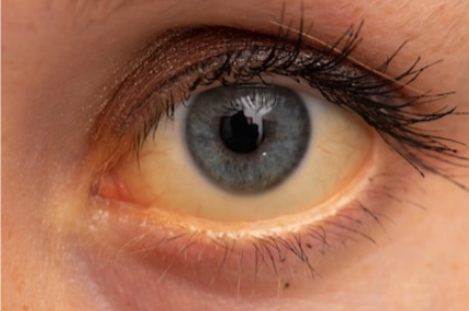
Yellow Whites of Your Eyes

When your skin and also eyes look yellow, that’s called jaundice. It
usually implies you have liver troubles as well as is brought on by
high degrees of bilirubin, something your liver makes even more of
when it’s swollen or damaged. Poor diet regimen, cancerous cells,
infection, as well as persistent alcoholic abuse can all harm your
liver. Therapy varies from way of living adjustments to medicine to
liver transplants. Tiny yellow areas (not complete yellow) is triggered
from sunlight damages and also can be eliminated with a small
treatment.

Eye Twitches

Eye Twitches
Eye Twitches

These are very usual and also generally safe – they normally
disappear by themselves. They can be related to alcohol, exhaustion,
high levels of caffeine, insufficient rest, stress and anxiety, or
smoking cigarettes. In exceptionally uncommon instances, they can
be an indication of an issue with your nerve system, like multiple
sclerosis. Yet if the twitches are connected to MS or an additional
issue with your nerve system, you would certainly have various other
signs and symptoms, also, like trouble walking, speaking, and also
{going to the restroom.

Night Blindness

Night Blindness
Night Blindness

If it’s tough for you to see in low light, you may require glasses or you
might have cataracts – an all-natural component of ageing. Yet night
blindness is not usual amongst more youthful people in the United
States. In uncommon scenarios, you might not be getting sufficient
vitamin A. This is rather typical in poorer nations where diets may
not be sufficient to cover all the basic nutrients. It’s treated with
supplements or a diet plan with foods high in vitamin A, like sweet
potatoes, beef liver, spinach, other green leafy vegetables, carrots,
as well as pumpkins. An uncommon reason is also retinitis
pigmentosa.

Down syndrome and vision related issues

Down syndrome has effects on the developing eye which can affect the proper development of vision. Eye disease is seen to be reported in over fifty percent of patients with Down Syndrome, from less severe problems such as tear duct abnormalities to vision threatening diagnoses such as early age cataracts, which could severely affect vision. Specific focus ought to be given to vision & vision treatment in people with Down syndrome.

What is Different About the Eyes in People with Down Syndrome?

As any type of family member of a person with Down syndrome recognizes, there are particular features regarding the eyes, which are apparent visually. This includes upward slanting of the eyelids, prominent folds of skin in between the eye and the nose, as well as small white spots present on the iris (the tinted part of the eye) called Brush field’s spots. These spots are safe and not harmful, and can be seen in people without Down syndrome too.

Do The majority of Children with Down Syndrome Require Glasses?

These Refractive errors (the need for glasses is much more common in children with Down syndrome than in the general population) can be hyperopia (Farsightedness), astigmatism, or myopia (Near-sightedness). One more common problem is weak accommodation (which is problem in changing the focusing power of the eye from distance to near). We can evaluate this quickly in the clinic, and also if identified, we will certainly recommend glasses that have bifocals. Several of these types of patients have difficulty adapting to glasses, but once they get accustomed to having the glasses on their face, their vision is considerably better and usually their eye alignment improves as well.

What Are Common, however Less Serious, Eye Abnormalities Influencing Patients with Down Syndrome?

In addition to the demand for spectacles, lots of children with Down syndrome have tear duct abnormalities. Family members of the patient will observe this as a frequent discharge from the eyes and tear drop formation or watery eyes, intensified by colds. We usually recommend firm massage over the space between the eye and the nose tear region 2-3 times in a day to attempt to open up the tear duct. If this continues past a year without any improvement, the tear ducts might require to be opened up by a surgery.

Strabismus (eye imbalance) is also much more usual. Members of the family might discover that the eyes do not align well with each other but often the strabismus can be refined, which is even clear to the paediatrician. The folds of skin situated between the eyes and the nose can additionally conceal the underlying strabismus, or make the eyes appear as if they are crossing also, even when they are not. It is important to diagnose strabismus as a child, as crossed eyes can result in amblyopia (loss of vision which is also additionally known as lazy eye) and loss of stereopsis (using both eyes together), or depth perception.

How Exactly Can the Strabismus Be Treated?

In some cases, merely glasses alone are enough to align eyes with strabismus. If glasses are required, we usually begin there. If the eyes continue to have strabismus despite the appropriate pair of eyeglasses, after that we recommend strabismus surgical procedure (eye muscle surgery). This is a one to two hours procedure, which can often be done as an outpatient basis, unless there are various other reasons that the person would certainly require to be admitted, such as a serious heart condition. Unfortunately, our patients with Down syndrome are more likely to require more than one surgery to align their eyes as they don’t always react as predictably to strabismus surgical treatment as the general population with strabismus would certainly react.

What are the more severe eye problems that can develop unexpectedly?

My greatest concern is congenital cataracts (absence of clearness to the lens of the eye). If visually significant cataracts are present early in a child’s eye, after that a clear image is not delivered to the brain as well as therefore the brain can never “discover” to see. This is an extreme form of amblyopia referred to as derivational amblyopia. While we can take our time eliminating a cataract in a grown-up patient, substantial cataracts existing very early in a child’s life that are not removed can lead to lifelong poor inadequate vision. Because of circumstances, even if the cataract is eliminated when the child is older, the vision never improves significantly. This is what makes early detection of cataracts in babies and children so crucial. A child with Down syndrome will certainly be evaluated by the paediatrician at birth for any abnormalities or defects, as well as referred to an ophthalmologist if something abnormal is detected.

There is also a unique type of cataract in Down syndrome patients that we have actually discovered in our research study. However, depending upon how developmentally delayed the person is, they may not be able to communicate that they can’t see. For this reason, consequently, I recommend any type of patient with Down syndrome, regardless of age, have a complete eye examination if they are starting to show reduced cognitive function, or modifications in their regular activities.

Are There Various Other Eye Conditions in Down Syndrome That Can Cause Loss of Eyesight?

I mentioned amblyopia (commonly called “lazy eye” which is usually decreased vision) which can be triggered by multiple & various eye problems such as strabismus, severe ptosis (eyelid droop), cataracts, and even uncorrected refractive error, especially if one eye requires a much stronger eyeglass prescription than the other. Ptosis is usually easier to appreciate but strabismus and significant refractive error can be extremely challenging for the paediatrician to diagnose. There are various other much more unusual issues which can occur with the optic nerve or retina of the eye which can sometimes trigger vision loss and unfortunately are generally not treatable. Nystagmus (a balanced trembling of the eyes) can also occur.

What Are the Eye Care Recommendations for Children with Down Syndrome?

The American Academy of Paediatrics (AAP) and the Down Syndrome Medical Interest Group (DSMIG) recommend evaluation of the red response of the eyes at birth to search for cataracts, in addition to assess the eyes for strabismus or nystagmus. The red reflex is basically the “red eye” seen in photography, which is the regular response of the retina when struck by direct light. If the eyes don’t look normal, then the infant will be referred to a paediatric ophthalmologist—a medical professional who has completed specialized training in medical as well as surgical monitoring of the child’s eye. We, along with the AAP and the DSMIG, recommend a child with Down syndrome has their very first eye examination by an ophthalmologist experienced in patients with special disabilities (as an example, a paediatric ophthalmologist) by six months of age. After that, children with Down syndrome, even if they are without signs and symptoms, ought to see an ophthalmologist every one to two years. If any type of eye problem is detected, they will certainly be required to meet much more regularly.

What Sort of Symptoms We Might See If a Child Has an Eye Problem?

Unfortunately, children with Down Syndrome frequently do not complain about their eye problems, either due to the fact that they don’t observe the trouble or because they can’t communicate the problem well enough. Indications to search for include, squinting or closing one eye shut as a habit to be able to see well, an unusual head tilt, crossing or wandering of one or both eyes, or light sensitivity. In some severe cases, the sign of vision problems may be a regression in overall feature or loss of developmental milestones. Ptosis will certainly be seen as a cover droop, as well as a blocked tear duct will result in daily tearing and discharge.

Any Kind of Thoughts for Parents of a Child with Down Syndrome Who Are Worried About their Eye or Vision?

Obtaining normal eye examinations is very important in children with Down syndrome because eye disorders are so common and are challenging for the doctor to diagnose.  Since the examination can be challenging for both the child and also the doctor professional, it is best to have the examination done by an ophthalmologist skilled in dealing with children with developmental delays. Don’t be surprised to find out your child’s needs for glasses; if needed, the glasses will certainly help the vision, and perhaps the eye positioning also, along with helping in the development of normal vision pathways in the brain. This will certainly help with your child’s understanding as well as functioning. Our research is looking at simply exactly how usual eye problems are in Down syndrome, in addition to the growth of cataracts in these patients.

Vision loss after stroke: Reality sheet

Vision loss after stroke: Reality sheet
Don’t Drive After Surgery Says Beverly Hills Ophthalmologist

About one-third of stroke survivors experience vision loss.

· Many people who have vision loss after a stroke do not fully recover their vision.

· Some recuperation is possible– this will generally happen in the very first couple of months after a stroke.

· Training, equipment and house modifications can assist you to live as independently and safely as possible.

Vision loss after stroke

Your vision depends on a healthy eye to receive information and healthy and balanced brain to process these details. The nerves in the eye travel from the eye through the brain to the occipital cortex at the back of the brain, enabling you to see.

The majority of strokes impact one side of the brain. Nerves from each eye traveling with each other in the brain, so both eyes are affected. If the right side of your brain is damaged, the left side vision in each eye might be affected. It is uncommon for both sides of the brain to be affected by stroke. When it does happen, it can lead to blindness.

TYPES OF VISION LOSS

Visual field loss

Your visual field is the entire area you can see when your eyes are dealt with in one position.

Homonymous hemianopia is the loss of fifty percent of the visual field in each eye. You may feel like you are unable to see out of one eye, as you see only half images, however actually, both your eyes are affected. When reading, words and sentences disappear when in the missing visual field as they cannot be seen. People may appear to have only half a face.

Quadrantanopia is the loss of either the upper or lower quarter of the visual field, therefore the name derived from quadrant or quarter of a circle.

Eye movement control

If the nerves that make your eyes move are harmed, you may not be able to move your eyes to a specific position in order to see objects that lie in that position. Another effect may be that a nerve that controls individual eye muscles may stop working, triggering eye turning (technically called strabismus) or double vision (technically called diplopia). Various other problems with eye nerves can create an eyelid to droop (ptosis), or the pupil of an eye to come to be bigger.

Unstable movement

Nystagmus is constant, unstable movement of the eyes. Activity will certainly be jittery and can be side to side, backwards and forwards or circular. Your eyes may also flicker when you try to look continuously at something.

Dry eyes

Troubles with the nerves of the eyelid, the facial nerve or the muscles of the eyelid can cause dry eyes.

Visual overlook and agnosia

People with visual neglect are not familiar with, and do not react to, things on their stroke-affected side. People with visual agnosia have trouble identifying familiar faces as well as objects. These problems are not associated with vision itself, though that may also be affected – they result from damages to parts of the brain that perceive and interpret vision or the visual images that are formed in the eye.

Management and recovery

The majority of people that have vision loss after a stroke will certainly not completely recover their vision. Some recuperation is possible, usually in the very first couple of months after a stroke. Glasses or contact lenses generally will not help vision loss due to stroke.

Ophthalmologists, orthoptists, medical professionals and occupational therapists can recommend on the very best monitoring for you.

Treatment may include training to compensate for the vision loss. This consists of scanning techniques that make the most use of your existing vision.

Prism lenses can be used to move images from the non-seeing to the seeing visual field. If you have dual vision, a prism to sign up with the images can be positioned on glasses, or an eye spot can assist by blocking one image.

You can utilize eye drops for dry eyes. Your health and wellness expert may also recommend taping your eyelids shut, especially during the night. If you have become more sensitive to light (photo sensitivity), you can use sunglasses.

Vision loss can affect your safety and independence. Training, aids, equipment and modifications can help you to lead close to a normal life.

More help

Khanna Vision Institute’s health and wellness professionals provide information, guidance, support and recommendation.